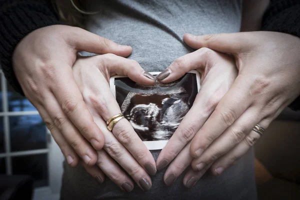
Ultrasound technology has been used for decades to monitor the development of fetuses during pregnancy. With advancements in technology, 3D ultrasounds have become increasingly popular among expecting parents.
These ultrasounds provide a more detailed view of the baby’s features, allowing parents to see their child’s face, fingers, and toes. However, many parents wonder, ‘Can You See Hair on 3D Ultrasound?’
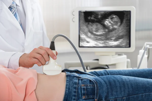
Before discussing whether hair is visible on a 3D ultrasound, it’s important to understand how this technology works. Unlike traditional 2D ultrasounds, which provide a flat image of the baby, 3D ultrasounds use sound waves to create a three-dimensional image of the baby.
This technology provides a more detailed view of the baby’s features, including facial features, fingers, and toes.
While 3D ultrasounds provide a more detailed view of the baby’s features, it’s important to note that hair may not always be visible. Hair is typically visible on a 3D ultrasound after 25 weeks of gestation.
However, this can vary depending on the baby’s position, the amount of amniotic fluid, and other factors. It’s also important to note that the quality of the ultrasound machine and the skill of the technician can affect the visibility of hair on a 3D ultrasound.
Key Takeaways
- 3D ultrasounds provide a more detailed view of the baby’s features, but hair may not always be visible.
- Hair is typically visible on a 3D ultrasound after 25 weeks of gestation, but this can vary depending on various factors.
- The quality of the ultrasound machine and the skill of the technician can affect the visibility of hair on a 3D ultrasound.
1. Understanding 3D Ultrasound

3D ultrasound is a medical imaging technique that generates three-dimensional images of the fetus in the womb. Unlike traditional 2D ultrasound, which produces flat, two-dimensional images, 3D ultrasound uses sound waves to create a three-dimensional image of the fetus in the womb.
The ultrasound machine sends high-frequency sound waves into the body, which bounce back off the fetus and surrounding tissues to create an image. The sound waves are then converted into a three-dimensional image by a computer, which can be viewed on a monitor.
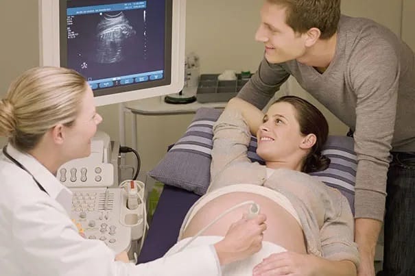
3D ultrasound offers several advantages over traditional 2D ultrasound. It provides a more detailed view of the fetus and can help detect abnormalities that may not be visible in 2D ultrasound.
It also allows parents to see their baby’s features and movements in greater detail, which can be a very emotional and rewarding experience.
However, it is important to note that 3D ultrasound is not always necessary or recommended for every pregnancy. It is typically used in cases where there may be a concern about the baby’s health or development, or in cases where parents want a more detailed view of their baby.
In terms of hair, it is possible to see hair on a 3D ultrasound image, although it may not always be visible depending on the position of the baby and the quality of the image. It is also important to note that hair on a fetus is not always a reliable indicator of what the baby’s hair will look like after birth, as hair can change color and texture in the weeks and months after birth.
Overall, 3D ultrasound is a valuable tool in prenatal care, providing a more detailed and emotional view of the developing fetus. However, it is important to use it appropriately and in conjunction with other prenatal tests and screenings to ensure the health and well-being of both the mother and the baby.
2. Hair Visibility in 3D Ultrasound

One common question that many expectant mothers have is whether they can see their baby’s hair on a 3D ultrasound. While it is possible to see hair on a 2D ultrasound, the visibility of hair on a 3D ultrasound can vary.
In general, hair on a 3D ultrasound may be visible if the baby has a significant amount of hair. However, the visibility of individual strands of hair can be difficult to determine, as the resolution of the ultrasound may not be high enough to show fine details.
Additionally, the color of the hair can also affect its visibility on a 3D ultrasound. White strands of hair may be easier to see than darker strands, as they reflect more light and create a stronger contrast against the baby’s skin.
It is also important to note that not all babies have hair visible on a 3D ultrasound. Hair growth can vary from baby to baby, and some babies may not have enough hair to be visible on the ultrasound.
Overall, while it is possible to see hair on a 3D ultrasound, the visibility of hair strands and follicles can vary depending on factors such as hair thickness, color, and growth.
3. Hair Growth During Pregnancy

Hair growth is a common phenomenon during pregnancy, and it is a result of hormonal changes in the body. The hair on the head and other parts of the body may grow faster and thicker, while some women may experience hair loss during pregnancy.
During the first trimester, the baby’s hair starts to grow in the form of fine, soft hair called lanugo. This hair covers the entire body of the fetus and helps regulate the body temperature.
The lanugo hair is usually shed during the third trimester and replaced by terminal hair, which is the hair that grows on the scalp, eyebrows, and eyelashes.
The hair on the scalp of the fetus grows at a rate of about 0.5 mm per day during the third trimester. The hair growth is influenced by the hormones produced by the mother, including estrogen and progesterone.
These hormones stimulate the hair follicles and promote hair growth.
The hair growth on the fetus is not limited to the scalp. The fetus also develops hair on other parts of the body, such as the back, arms, and legs.
This hair is called fetal hair and is usually shed shortly after birth.
It is important to note that not all fetuses develop the same amount of hair. Some may have more hair than others, and some may not have any hair at all.
The amount of hair growth is determined by various factors, including genetics and the stage of fetal development.
In conclusion, hair growth during pregnancy is a normal and natural process that is influenced by hormonal changes in the body. The fetus develops hair on various parts of the body, including the scalp, and this hair is shed and replaced by terminal hair during the third trimester.
4. Interpreting Ultrasound Images

Interpreting ultrasound images can be challenging, especially for those who are not familiar with the technology. Ultrasound images are typically black and white, and they can be difficult to interpret without the proper training.
However, with a little knowledge and practice, it is possible to read and understand these images.
When looking at an ultrasound image, there are several things to keep in mind. First, pay attention to the texture of the image.
Areas that are smooth and uniform in texture are likely to be fluid-filled, while areas that are more irregular in texture may indicate the presence of solid tissue.
Another important factor to consider when interpreting ultrasound images is the presence of shadows. Shadows can be caused by the reflection of sound waves off of solid objects, such as bones or organs.
These shadows can help to identify the location and shape of these structures.
Contours are also an important aspect of interpreting ultrasound images. The contours of organs and tissues can provide valuable information about their size, shape, and location.
In addition, the contours of blood vessels can help to identify areas of blockage or narrowing.
One common feature of ultrasound images is the presence of a fuzzy halo around objects. This halo is caused by the scattering of sound waves as they pass through different tissues.
While this halo can make it difficult to see fine details, it can also provide important information about the overall shape and size of objects.
Overall, interpreting ultrasound images requires a combination of knowledge and practice. By paying attention to the texture, shadows, contours, and fuzzy halos of ultrasound images, it is possible to gain a better understanding of the structures and tissues that are being imaged.
5. Role of Genetics in Hair Growth

Hair growth is a complex process that is influenced by various factors, including genetics. The genes we inherit from our parents play a crucial role in determining the thickness, texture, and color of our hair.
Babies’ hair growth is also influenced by genetics. Some babies are born with a full head of hair, while others have very little hair at birth.
The amount of hair a baby has at birth is not an indication of how much hair they will have later in life.
The color of a baby’s hair is also determined by genetics. Hair color is determined by the amount and type of melanin in the hair shaft. Melanin is a pigment that gives hair its color.
Babies with more melanin will have darker hair, while those with less melanin will have lighter hair.
Family history can also play a role in determining hair growth patterns. If a baby’s parents or siblings have thick, curly hair, there is a higher chance that the baby will also have thick, curly hair.
In summary, genetics plays a significant role in hair growth, including baby hair and hair color. Family history can also provide clues about a baby’s hair growth patterns.
6. Additional Information on 4D Ultrasound
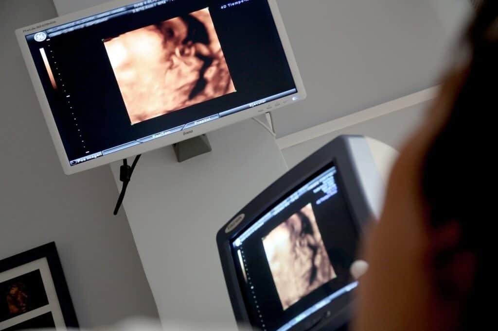
4D ultrasound is an advanced technology that allows doctors and parents to see a three-dimensional image of the baby in real-time. The fourth dimension is time, which means that the image is moving, and parents can see their baby’s movements and facial expressions.
HDlive 3D ultrasound is a type of 4D ultrasound that provides high-definition images of the baby. It uses advanced technology to produce realistic images that resemble a photograph.
This technology uses a combination of light and sound waves to create an image of the baby.
One of the benefits of 4D ultrasound is that it can detect potential problems with the baby’s development. Doctors can use 4D ultrasound to check for abnormalities in the baby’s organs, limbs, and other body parts.
It can also be used to monitor the baby’s growth and development.
Another benefit of 4D ultrasound is that it allows parents to bond with their baby before it is born. They can see their baby’s face and movements, which can be a very emotional and rewarding experience.
It is important to note that 4D ultrasound is not a replacement for regular prenatal care. It is a diagnostic tool that can provide additional information about the baby’s health and development.
Parents should always consult with their doctor about any concerns they have about their baby’s health.
In conclusion, 4D ultrasound, including HDlive 3D ultrasound, is an advanced technology that provides parents with a unique opportunity to see their baby in real-time. It can help detect potential problems with the baby’s development and allow parents to bond with their baby before it is born.
However, it is important to remember that 4D ultrasound is not a replacement for regular prenatal care and should always be used in conjunction with other diagnostic tools.
7. Understanding Abnormalities via Ultrasound

Ultrasound is a powerful tool for detecting abnormalities in the body. It can detect tumors, cysts, and other abnormalities that may not be visible on the surface.
Ultrasound is often used to diagnose and monitor the progress of these abnormalities.
A tumor is a mass of tissue that is abnormal in size, shape, or function. Tumors can be benign or malignant. Benign tumors are not cancerous and do not spread to other parts of the body.
Malignant tumors are cancerous and can spread to other parts of the body. Ultrasound can detect the presence of a tumor and determine its size and location.
Cysts are fluid-filled sacs that can form in any part of the body. They can be benign or malignant. Benign cysts are not cancerous and do not spread to other parts of the body.
Malignant cysts are cancerous and can spread to other parts of the body. Ultrasound can detect the presence of a cyst and determine its size and location.
Abnormalities can also be detected through ultrasound. These abnormalities can be caused by a variety of factors, such as genetic disorders, infections, or injuries.
Ultrasound can detect the presence of an abnormality and determine its location and severity.
It is important to note that ultrasound is not always 100% accurate in detecting abnormalities. Some abnormalities may be too small to be detected by ultrasound, while others may be obscured by other structures in the body.
In some cases, further testing may be necessary to confirm the presence of an abnormality.
In conclusion, ultrasound is a valuable tool for detecting and monitoring abnormalities in the body. It can detect tumors, cysts, and other abnormalities that may not be visible on the surface.
While it is not always 100% accurate, ultrasound can provide valuable information for diagnosis and treatment.
8. Ultrasounds and Body Fat

Ultrasounds are widely used in medical imaging to produce images of internal organs, tissues, and structures. However, the accuracy of ultrasound imaging can be affected by various factors, including the amount of body fat present in the patient.
Body fat can interfere with the ultrasound waves, making it difficult for the technician to obtain clear images. This is because ultrasound waves have a harder time penetrating fatty tissues compared to other types of tissues, such as muscle or bone.
Moreover, the amount of body fat can also affect the quality of the images obtained during the ultrasound. In general, the more body fat a person has, the less clear the images will be.
This is because the ultrasound waves are scattered and absorbed by the fatty tissues, resulting in a lower resolution and contrast of the images.
To address this issue, some ultrasound machines are equipped with special probes that can penetrate through the fatty tissues more effectively. Additionally, some techniques such as changing the patient’s position or using a different frequency of ultrasound waves can also help improve the quality of the images obtained.
In conclusion, body fat can affect the accuracy and quality of ultrasound imaging. Patients who have a higher amount of body fat may experience more difficulty in obtaining clear images during an ultrasound procedure.
However, with the use of specialized equipment and techniques, it is possible to overcome these challenges and obtain accurate and high-quality ultrasound images.
9. Expectations for Expecting Parents
When expecting parents go in for a 3D ultrasound, they may have certain expectations about what they will be able to see. It’s important to understand that while 3D ultrasounds can provide a more detailed look at an unborn baby, there are limitations to what can be seen.
One thing to keep in mind is that the quality of the images can vary depending on factors such as the position of the baby and the amount of amniotic fluid. This means that some images may be clearer than others, and it may not always be possible to get a perfect view of the baby’s features.
Another expectation that some parents may have is that they will be able to see the baby’s hair on a 3D ultrasound. While it is possible to see hair on an ultrasound, it’s important to understand that this is not always the case.
The baby’s hair may not be visible if it is not long enough or if it is not in a position that allows it to be seen.
Overall, it’s important for expecting parents to have realistic expectations when it comes to what they will be able to see on a 3D ultrasound. While these images can provide a more detailed look at an unborn baby, there are limitations to what can be seen.
It’s best to focus on enjoying the experience of seeing the baby and getting a glimpse of what he or she will look like when born.
10. Hair and Premature Babies
One of the most common questions asked by expectant mothers is whether or not they will be able to see their baby’s hair on a 3D ultrasound. While it is possible to see hair on a 3D ultrasound, it is not always clear, especially in premature babies.
Premature babies, also known as pre-term babies, are born before 37 weeks of gestation. These babies are often smaller and less developed than full-term babies, which can make it more difficult to see certain features on a 3D ultrasound, including hair.
While premature babies may have some hair, it is often very fine and light in color, making it difficult to see on a 3D ultrasound. Additionally, premature babies may have less hair than full-term babies, as hair growth is one of the last things to develop in utero.
It is important to note that the ability to see hair on a 3D ultrasound can vary depending on the quality of the ultrasound machine and the skill of the technician performing the ultrasound. In some cases, it may be possible to see more detail, including hair, on a higher quality machine or with a more experienced technician.
Overall, while it is possible to see hair on a 3D ultrasound, it may not always be clear or visible, especially in premature babies. It is important to remember that the purpose of a 3D ultrasound is not to determine the amount of hair on a baby’s head, but rather to monitor the baby’s growth and development.
11. Myths Related to Hair and Heartburn
There are many myths surrounding pregnancy, and two of the most common ones are related to hair and heartburn. Many people believe that if a pregnant woman experiences heartburn, it means that her baby will have a full head of hair. However, this is not necessarily true.
Studies have shown that there is no correlation between heartburn and the amount of hair a baby has. While heartburn is a common symptom of pregnancy, it is caused by the hormone progesterone, which relaxes the muscles in the esophagus and allows stomach acid to flow back up into the throat.
This has nothing to do with the amount of hair on a baby’s head.
Another myth is that if a pregnant woman has heartburn, it means that her baby will be born with a lot of hair. However, this is also not true.
The amount of hair a baby has is determined by genetics, not by the mother’s heartburn.
In conclusion, while heartburn is a common symptom of pregnancy, it is not related to the amount of hair a baby has. The amount of hair a baby has is determined by genetics, and there is no way to predict how much hair a baby will have based on the mother’s heartburn.
12. Determining Baby’s Gender

Determining a baby’s gender is one of the most exciting parts of pregnancy for many parents. While there are many old wives’ tales and myths about how to predict the sex of a baby, a 3D ultrasound is one of the most reliable ways to determine the gender of a baby before birth.
During a 3D ultrasound, the sonographer will be able to see the baby’s genital area and determine whether they are male or female. This is done by looking for the presence of a penis or labia.
In some cases, it may be difficult to determine the gender of the baby, especially if they are in a position that makes it hard to see their genital area.
It is important to note that while a 3D ultrasound is a reliable way to determine the gender of a baby, it is not always 100% accurate. In some cases, the sonographer may mistake the genital area for something else, or the baby’s position may make it hard to see their genital area clearly.
Overall, a 3D ultrasound is a great way to determine the gender of a baby before birth. However, parents should keep in mind that the accuracy of the ultrasound may vary and that it is always possible for the sonographer to make a mistake.
Related post: What Does Hair Look Like on an Ultrasound
Frequently Asked Questions
What can 3D ultrasound detect?
3D ultrasound is a medical imaging technique that uses sound waves to create a three-dimensional image of the baby in the womb. It can detect various aspects of the baby’s development, including the size and shape of the head, limbs, and organs. It is also used to check for any abnormalities in the baby’s development.
Can you tell if the baby has hair in a 3D ultrasound?
Yes, it is possible to see if the baby has hair in a 3D ultrasound. However, the quality of the image can vary depending on factors such as the baby’s position, the amount of amniotic fluid, and the mother’s body size. Some 3D ultrasounds may show more detail than others.
What does a lot of hair look like on ultrasound?
A lot of hair on ultrasound may appear as a fuzzy or hazy area on the baby’s head. It may also appear as a dark area on the ultrasound image.
Why do 3D ultrasounds look lumpy?
3D ultrasounds can sometimes look lumpy due to the way the images are created. The ultrasound machine takes multiple images of the baby from different angles and then combines them to create a 3D image. The resulting image can sometimes appear lumpy or distorted.
Can you tell if a baby has hair on a 5D ultrasound?
There is no such thing as a 5D ultrasound. The term “5D ultrasound” is a marketing term used by some ultrasound providers to refer to a 3D ultrasound with added features such as color and movement. It is still possible to see if the baby has hair on a 3D ultrasound with added features.
How early can you see hair on an ultrasound?
Hair can start growing on a baby’s head as early as 14 weeks gestation. However, it may not be visible on an ultrasound until later in the pregnancy when the baby’s hair is longer and thicker. The quality of the ultrasound image can also affect the visibility of the baby’s hair.

Iesha is a loving mother of 2 beautiful children. She’s an active parent who enjoys indoor and outdoor adventures with her family. Her mission is to share practical and realistic parenting advice to help the parenting community becoming stronger.