Hair plays an important role in our appearance and self-image. From the moment a baby is born, observers often comment on whether the newborn has a full head of hair or not. Expectant parents may wonder, “Can you see hair on ultrasound?”
This article will discuss the topic and delve into the key aspects related to hair and ultrasounds.
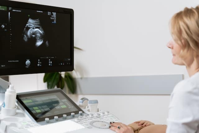
Ultrasounds are a widely used diagnostic tool during pregnancy, enabling doctors and sonographers to monitor the development of the fetus. They use high-frequency sound waves to create images of the fetus, allowing medical professionals to identify any potential concerns, measure growth, and prepare parents for what to expect.
While ultrasounds can provide a wealth of information about a developing fetus, it’s important to understand the limitations of the technology and to dispel any myths surrounding its capabilities.
Though ultrasounds may not provide a clear image of hair, there are insights to be gained about a baby’s potential hair growth and other factors.
By examining the science behind ultrasounds, genetics, and fetal hair development, expectant parents can better understand their baby’s development and set realistic expectations for what ultrasound images may show.
Key Takeaways
- Ultrasounds provide valuable information about fetal development, but their ability to show hair is limited.
- Genetics play a significant role in determining hair growth in unborn babies, along with other factors.
- It is essential to understand the limitations of ultrasound technology and to dispel myths associated with it.
Understanding Ultrasounds

Ultrasound technology uses sound waves to create images of the inside of the body. It is a non-invasive diagnostic tool that allows medical professionals to examine organs, blood vessels, and other internal structures.
This section will focus on the properties of sound waves, and the different types of ultrasounds, including 2D, 3D, and 4D imaging.
Properties of Sound Waves
Sound waves are mechanical waves produced by vibrating particles in a medium such as air or water. They travel in a longitudinal pattern, meaning that the medium’s particles move parallel to the direction of the wave.
In ultrasound imaging, high-frequency sound waves, typically between 2 and 18 MHz, penetrate tissue and reflect off boundaries between different structures. These reflected waves are then captured and analyzed to create an image, known as a sonogram.
Speed and attenuation: The speed of sound waves can affect the quality of the ultrasound image. Generally, sound waves travel faster in denser materials and slower in less dense materials.
The attenuation of the sound waves, or how they are absorbed by the tissue, also influences the image quality.
Frequency: The frequency of an ultrasound wave is directly related to its wavelength and is essential in determining the resolution of the image. Higher frequencies produce better resolution but have less penetration depth, making them more suitable for imaging superficial structures.
Lower frequencies facilitate deeper penetration but produce less detailed images.
Different Types of Ultrasounds
There are several types of ultrasounds, each with its own advantages and applications:
2D ultrasound: The most common type of ultrasound, 2D imaging, provides a flat, gray-scale picture of the internal structures. It is often used for fetal imaging, abdominal exams, and vascular imaging.
Its primary advantage is that it provides real-time images and can detect motion, including blood flow and fetal movements.
3D ultrasound: This type of ultrasound uses multiple 2D images to create a three-dimensional representation of the region being examined.
This technique allows for better visualization of the morphology and spatial relationships between structures, which can be helpful in prenatal examinations or diagnosing abnormalities.
4D ultrasound: Sometimes referred to as “real-time 3D,” 4D ultrasound is essentially a series of 3D images captured in rapid succession. This allows for real-time display of movement, such as a fetus kicking or waving its arms, making it particularly popular for prenatal imaging.
Overall, ultrasound technology offers a wide range of applications and imaging modes, each with specific advantages and use cases. By understanding how sound waves interact with tissues and the characteristics of various ultrasound types, medical professionals can utilize this versatile diagnostic tool effectively and safely.
Ultrasounds During Pregnancy
Determining the Baby’s Development
Ultrasounds are a useful tool during pregnancy, as they allow medical professionals to observe the baby’s development and assess its overall health. Throughout gestation, a series of ultrasounds may be conducted to monitor fetal growth and identify any abnormalities.
These scans will help to establish a more accurate due date and track critical aspects of fetal development, such as heartbeat, limb growth, and organ formation.
In the early stages of pregnancy, an ultrasound image may not show specific details of the baby, including the presence of fetal hair growth. However, as gestation progresses, more visible details and features of the baby can be observed, such as the presence of soft body hair called lanugo.
Monitoring Fetal Health
During pregnancy, it’s crucial to keep a close eye on the baby’s overall health and well-being. Ultrasounds are a non-invasive method for monitoring various aspects of the baby’s development, including:
- Growth rate: The ultrasound can determine if the baby is growing at a healthy pace or if there may be any underlying issues.
- Location of the placenta: The position of the placenta is essential as it can affect the delivery and the baby’s oxygen supply.
- Amniotic fluid levels: Adequate amniotic fluid is vital to cushion and protect the baby during pregnancy.
As the pregnancy progresses, ultrasounds can also be used to identify possible abnormalities in the baby’s development. Some of these issues can be related to hair growth. For example, babies with certain genetic disorders may have an excessive amount of hair or be born without hair.
While ultrasounds may not be the primary method for detecting hair-related issues, they can be helpful in identifying certain physical markers that may indicate an underlying issue.
In conclusion, ultrasounds during pregnancy contribute to determining the baby’s development and monitoring fetal health. While they may not provide an explicitly clear image of hair growth, they serve a crucial role in ensuring that both the mother and baby are progressing healthily throughout gestation.
Identifying Hair in Ultrasounds
Characteristic Appearance of Hair
In ultrasound imaging, hair may appear as white strands or a halo around the baby’s head. This is because hair on an ultrasound often reflects the sound waves, creating a distinct image.
While it can be challenging to identify individual strands of hair, it is possible to detect a full head of hair on a baby in certain cases. However, it’s important to note that the visibility of baby’s hair is highly dependent on the factors that influence ultrasound image quality.
Factors Influencing Visibility
There are several factors that can affect the visibility of hair on an ultrasound image. These include:
- Hair color: Hair color can influence how well hair appears on an ultrasound image. Darker hair generally reflects sound waves better than lighter hair, making it more visible.
- Ultrasound settings: The settings used during an ultrasound can have an impact on the clarity and detail of the image. Higher resolution and a focused area on the baby’s head can sometimes improve the chances of visualizing hair.
- Fetal position: The position of the baby inside the womb also plays a role in how well hair is displayed. If the baby is facing away from the ultrasound probe or their head is covered by other body parts, it may be difficult to see hair on the ultrasound.
- Gestational age: The gestational age of the baby can also influence the visibility of hair on an ultrasound. Fetal hair begins to grow around the 20th week of pregnancy and continues to develop throughout pregnancy. As the baby grows, it becomes more likely that hair can be detected in the ultrasound image.
In summary, identifying hair on an ultrasound can be challenging due to various factors that influence image quality. While it may be difficult to see individual strands, a full head of baby hair can sometimes be detected with the right conditions and settings.
Role of Genetics and Myths
Influence of Genetics on Hair Growth
Genetics play a significant role in determining hair growth and appearance. In fact, research from Johns Hopkins indicates that multiple genes are responsible for determining hair thickness, curliness, and color.
Moreover, these genes are inherited from both parents, allowing for a wide variety of hair types among individuals.
It’s important to note that ultrasound technology cannot detect hair follicles themselves. However, it can visualize the density of hair on a baby’s scalp, which is influenced by their genetics.
Old Wives’ Tales Explained
There are numerous old wives’ tales surrounding the correlation between fetal hair growth and the experiences of the mother during pregnancy. For instance, it’s widely believed that a mother experiencing heartburn is more likely to give birth to a baby with a lot of hair.
However, these myths have been debunked by scientific research.
Despite the prevalence of such tales, it’s crucial to remember that hair growth and appearance are determined by a combination of genetic factors, rather than external circumstances. Relying on folklore may be entertaining but isn’t a reliable method for predicting a child’s hair development.
In conclusion, ultrasound technology may provide a glimpse into a fetus’s hair density, but it’s essential to recognize the influence of genetics on hair growth and set aside age-old myths related to pregnancy experiences.
Details of Fetal Hair

Different Types of Hair
Fetal hair develops in distinct types during pregnancy. The primary type is lanugo, which appears around the fourth month of pregnancy. Lanugo is thin, soft, and lightly pigmented and covers the fetus’s body.
This coat of fine hair diminishes during the third trimester, and the fetus gradually replaces it with vellus hair, also known as peach fuzz. Vellus hair is short, fine, and less pigmented than other types of hair.
After birth, terminal hair, which is thick, strong, and darker in color, begins to replace vellus hair across different areas of the body.
Growth Patterns and Texture
Fetal hair growth patterns begin with the development of hair follicles on the fetus’s scalp around the 14th week of pregnancy. By the 20th week, the fetus’s entire body is covered in lanugo.
The growth and thickness of fetal hair vary between individuals, with some babies born with a full head of hair while others have very little or none at all. Hair texture in the womb can also range from fine to coarse, and straight to curly, with genetic factors playing a significant role in determining the eventual hair characteristics.
Role in In-Utero Development
Fetal hair, especially lanugo, plays essential roles in a baby’s development within the womb. Lanugo acts as insulation, keeping the fetus warm and protected as it develops in the uterine environment.
Additionally, lanugo helps anchor the protective layer of vernix caseosa, a waxy substance that shields the fetus’s skin from the surrounding amniotic fluid. The combination of lanugo and vernix caseosa also aids in babies’ smooth passage through the birth canal during delivery.
FAQs on Hair
- Can you see hair on ultrasound? While ultrasounds provide detailed images of a developing fetus, identifying specific hair types or patterns is generally challenging through ultrasound technology. However, in some cases, experienced sonographers may be able to detect a general presence or absence of hair, particularly on the scalp.
- Do all babies lose their lanugo after birth? Most newborns will shed their lanugo before or shortly after birth. However, some babies, particularly those who are born prematurely, might retain their lanugo for a more extended period before it gradually falls off.
- Does fetal hair color indicate the child’s future hair color? The color of the fetal hair may not necessarily indicate the child’s future hair color, as hair color can change over time due to various factors, including genetic influences and environmental factors.
Miscellaneous Topics
Potential Abnormalities
Ultrasound can detect potential abnormalities such as cysts and tumors in the developing baby. During the second and third trimester, sonographers can monitor the growth and development of the baby, checking for any signs of abnormalities.
It is important to note that ultrasounds are not 100% accurate in detecting all issues, but they are a valuable tool in identifying potential concerns.
Although hair cannot be seen on ultrasound, certain scalp abnormalities that affect the hair may be visible. For example, a fuzzy white halo could potentially signal a premature baby with underdeveloped hair follicles.
Identifying Gender
While ultrasound is not primarily used to determine the baby’s hair, it is often employed during the second trimester for gender identification. Around this period, the developing baby’s genitals become more distinguishable, allowing the sonographer to determine the gender with greater accuracy.
However, it is important to keep in mind that gender determination via ultrasound is not always correct due to factors such as the baby’s position and the quality of the ultrasound image.
Community Support and Resources
Expecting parents often experience a range of emotions and may face various challenges during the pregnancy stages. The community provides support systems and resources to help alleviate some of these challenges.
From baby products to registry builder tools, families can access numerous resources to prepare for their newborn’s arrival.
In addition to medical resources like morning sickness remedies and amniotic fluid testing, community support can be found in groups like prenatal classes and online forums where parents can share experiences and advice.
Furthermore, many people opt for in-utero photo shoots to capture early images of their developing baby, even though it might appear as a fuzzy blob on the screen.
The guidance and emotional support obtained from such communities can be invaluable for families during this crucial time in their lives, as they prepare to welcome a new member into their family.
Frequently Asked Questions
Is it possible to detect hair on a 3D ultrasound?
Yes, it is possible to detect hair on a 3D ultrasound. These advanced imaging techniques can show a more detailed view of the baby, including hair on the head, if it is present. However, it may not always be easy to distinguish between fine hair and other surrounding structures.
At what gestational age does hair become visible on an ultrasound?
Hair can become visible on an ultrasound as early as 20 weeks gestation. However, its visibility depends on factors such as the amount of hair, the quality of the ultrasound equipment used, and the skill of the technician performing the scan.
Does the ultrasound show if the baby has curly hair?
Ultrasound technology is not currently advanced enough to distinguish between different hair types, such as curly or straight hair. The images generated by ultrasounds focus on structural details, and differentiate between hair types is beyond their capabilities.
Can hair be seen during the 22-week ultrasound?
Hair may be visible during the 22-week ultrasound, as this is typically around the time when hair growth develops. However, as previously mentioned, the visibility of hair during a scan depends on various factors, including the hair density and the quality of the imaging technology.
How is the baby’s hair checked during a scan?
Baby’s hair is not a primary focus during routine ultrasounds, as medical professionals usually concentrate on essential features related to the baby’s health and development. If hair is observed during the scan, it might be a secondary finding.
The purpose of an ultrasound scan is to ensure the healthy development of the baby, not to examine hair.
Is hair visible on ultrasound at 36 weeks gestation?
At 36 weeks gestation, the visibility of hair through ultrasound is more likely than in earlier weeks. If the baby has a sufficient amount of hair, it might be visible on the ultrasound – primarily in 3D or 4D images.
However, keep in mind that the primary focus of these scans is still on the well-being of the baby and not the hair visibility.

Iesha is a loving mother of 2 beautiful children. She’s an active parent who enjoys indoor and outdoor adventures with her family. Her mission is to share practical and realistic parenting advice to help the parenting community becoming stronger.